
Visualizing Cellular Signaling in 2D Cell Culture Systems
We capture cellular signaling and spatial organization in 2D cell culture systems, including primary cells, immortalized lines, and co-cultures. Explore example images below from selected cell models we routinely work with.
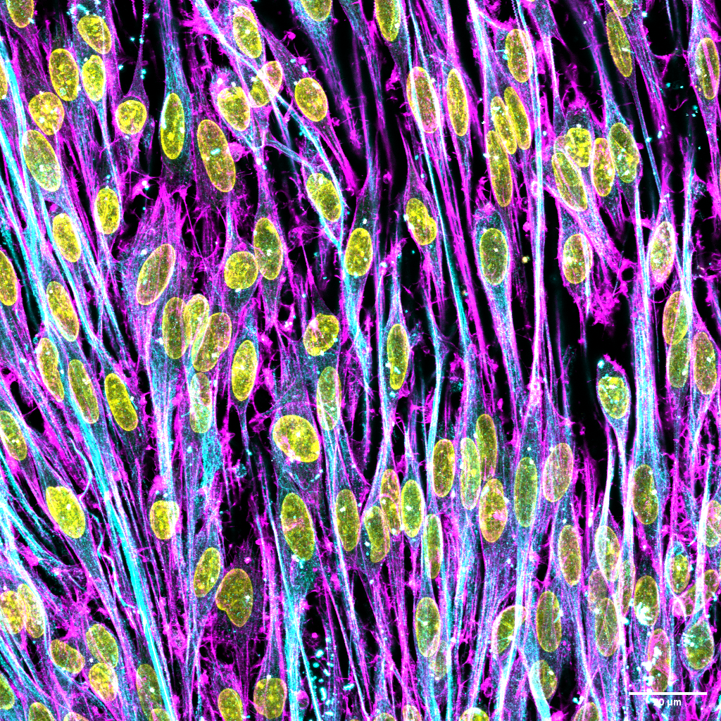
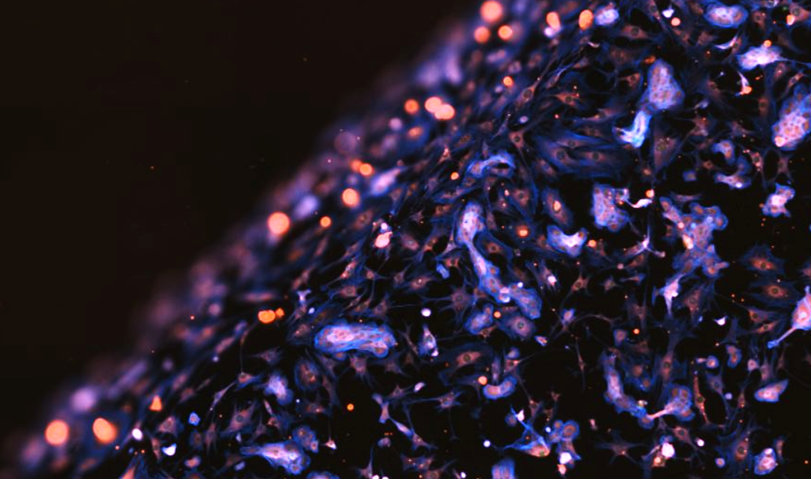
Murine Schwann cells on the surfaces of hydrogels
Markers: Acetylated tubulin, DAPI (nuclei) , F-actin (cytoskeletal structure)
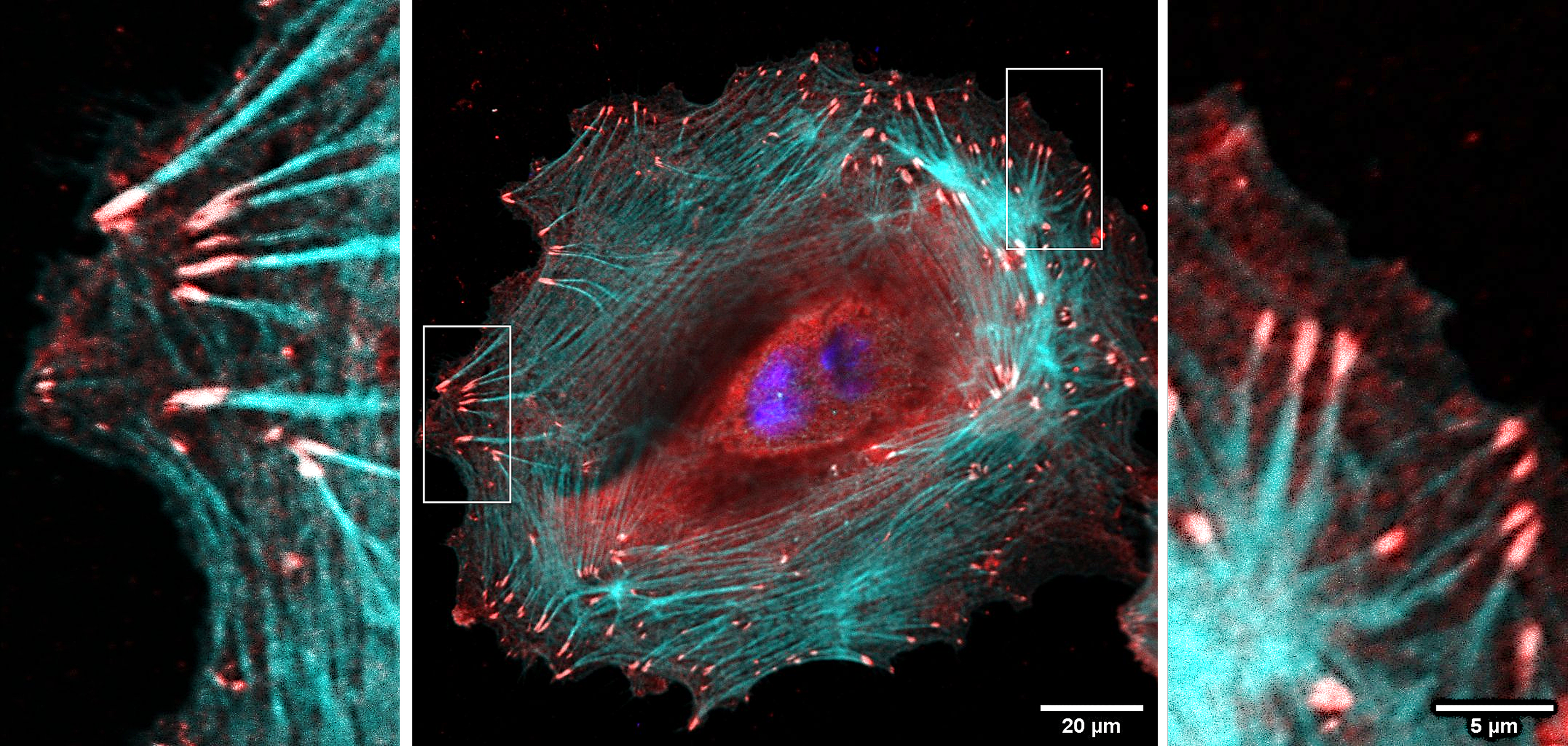
Human keratinocytes on a 2D biointerface
Markers: F-actin (cytoskeletal structure), vinculin (a key component of focal adhesions), DAPI (nuclei)
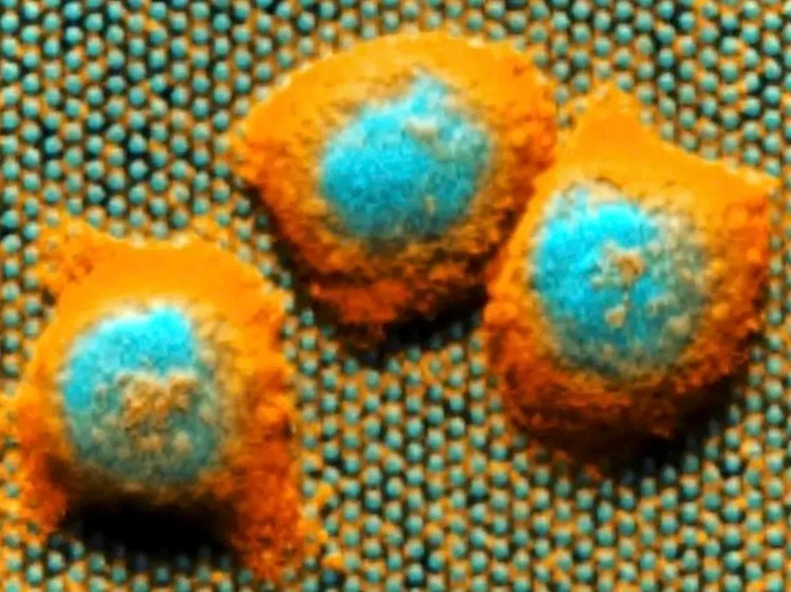
Murine Embryonic Stem Cells (mESCs) on nanostructures
Markers: F-actin (cytoskeletal structure), DAPI (nuclei)

Murine Schwann cells on protein-coated 2D surfaces
Markers: Acetylated tubulin, F-actin (cytoskeletal structure), DAPI (nuclei)
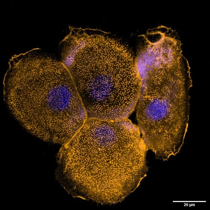
Human keratinocyte on protein-patterned surfaces
Markers: Alpha 6 integrin, DAPI (nuclei)
Murine Embryonic Stem Cells (mESCs) micro topographies
Markers: DAPI (nuclei), F-actin (cytoskeletal structure)