Visualizing Cellular Signaling

Representative ICC/IF examples across various biological systems.

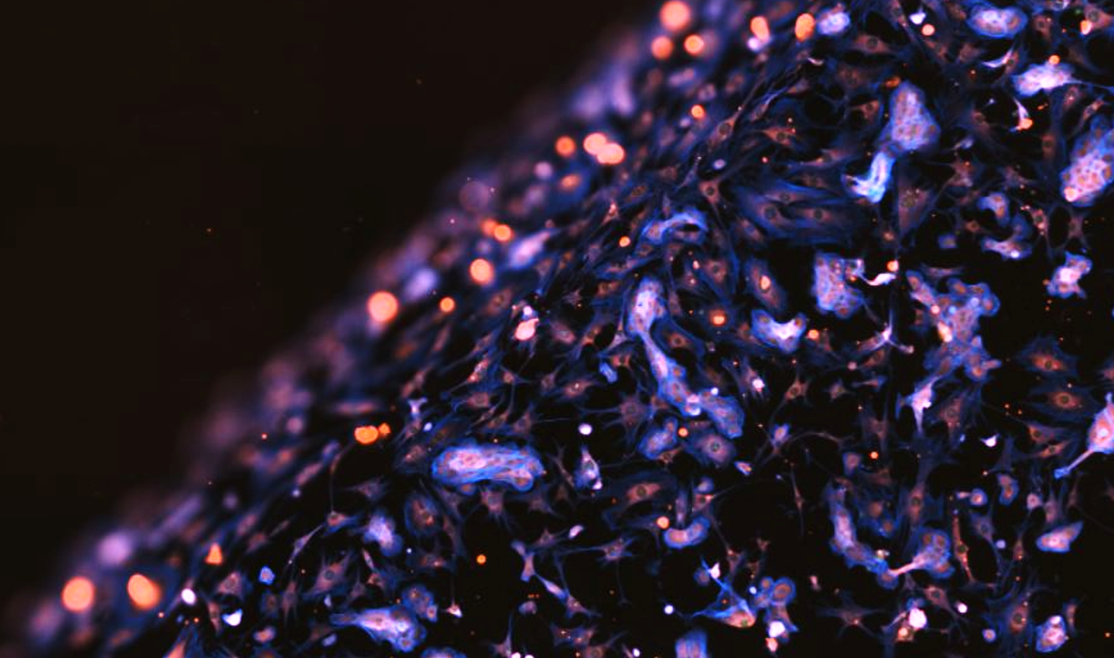
3D cell culture systems


Colorful microscopic view of neurons with neurite- ICC- βIII-Tubulin: marking neurite extensions
 🔴 F-actin: cytoskeletal structure
 🔵 DAPI: nuclear staining, #NeuralProgenitorCells hashtag#Neurodevelopment hashtag#NeuralDifferentiation hashtag#Neu

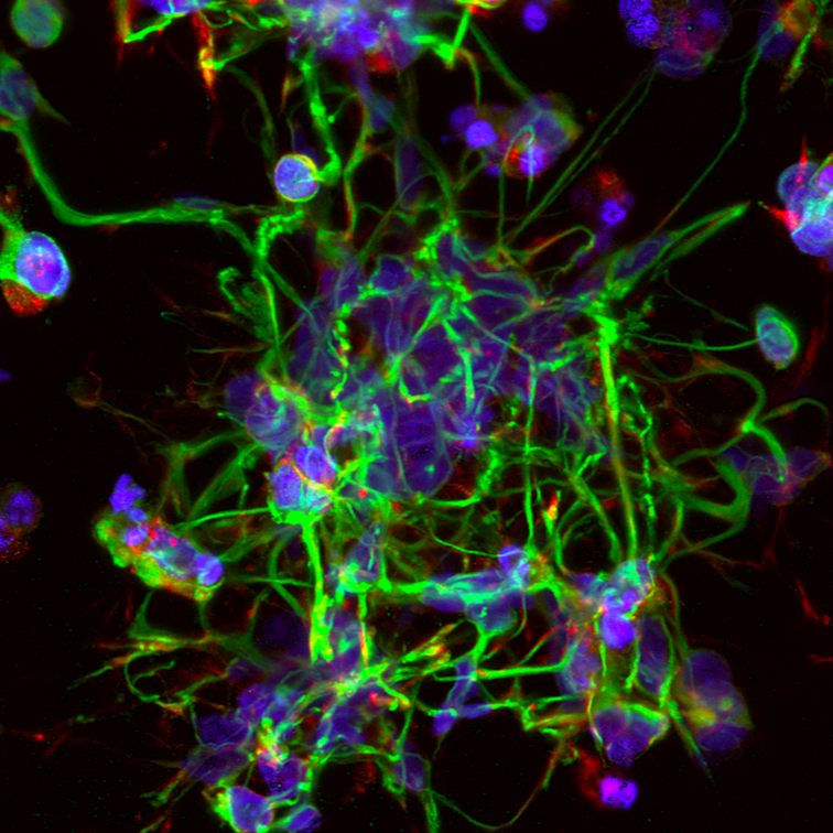
Human neural progenitor cells (NPCs) in 3D hydrogels

Nuclei (DAPI) human#endothelial cells cultured within a#bioprinted artificial#vascular system
 🟢 F-actin (Phalloidin, cytoskeleton)
 🔵 VE‑cadherin (endothelial junctions)#EndothelialCells#Bioprinting

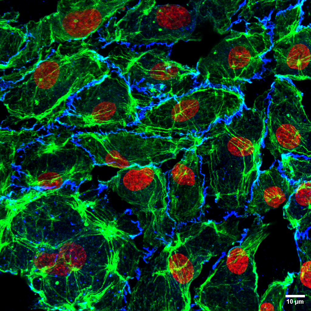
Human endothelial cells on 3D printed biomaterials

Brain#tumors remain among the most challenging cancers to treat, with#glioblastoma (GBM).Captured via hashtag#fluorescence microscopy, the image shows:
🟢 Collagen: a key component of ECM
🟣 Nuclei

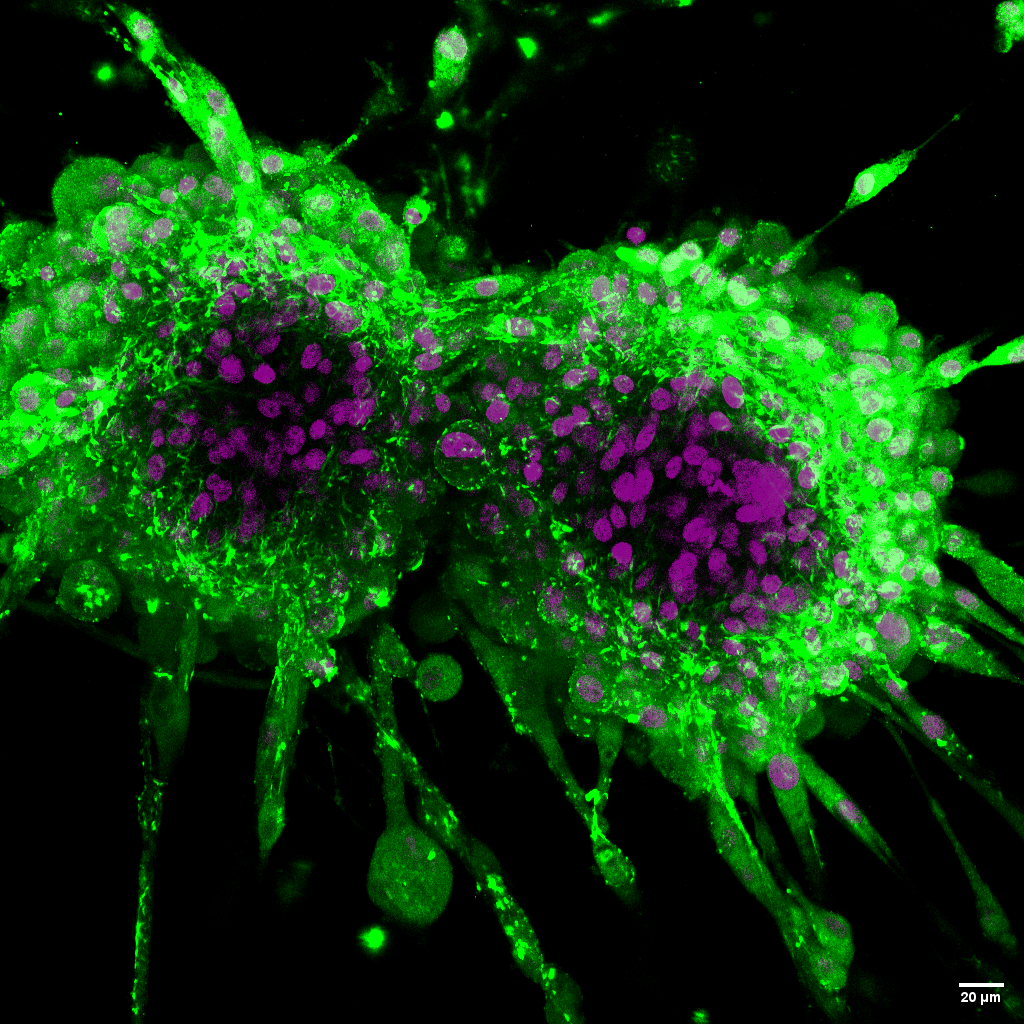
Glioblastoma cells in 3D hydrogels

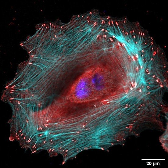
2D cell culture systems


#Schwann cells cultured on hashtag#biomimetic substrates designed to support peripheral nerve repair. Markers used in this#fluorescence image:
🟣 Acetylated Tubulin 
🟡 Nuclei 
🔵 F-actin

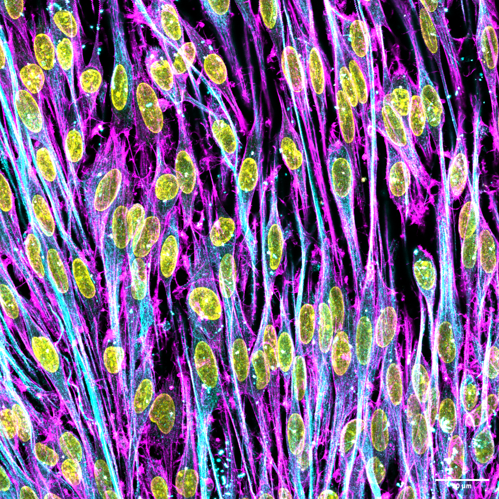
Murine Schwann cells on the surfaces of hydrogels

This week, we’re showcasing human hashtag#keratinocytes cultured on an#engineered cell-instructive surface designed to probe cell-matrix interactions.
Using#fluorescent labeling, we visualized:
🔴 Vinculin: a key component of focal a

Human keratinocytes on a 2D biointerface

Close-up of three tiny, spherical ESCs with orange and blue coloring, on a nanosctructures surface.

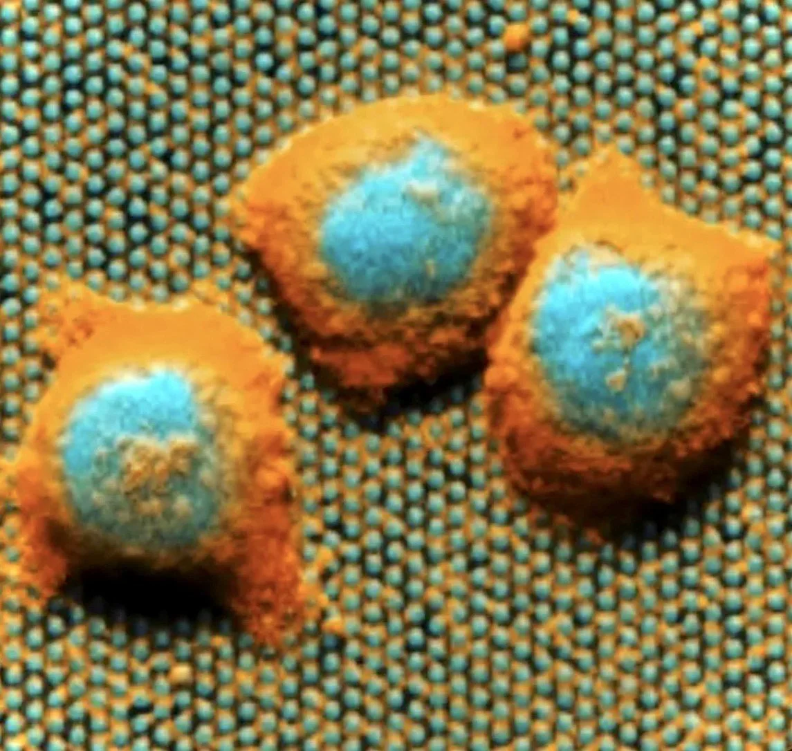
Murine Embryonic Stem Cells (mESCs) on the surface of nanostructures

Murine Schwann cells on protein-coated 2D surfaces

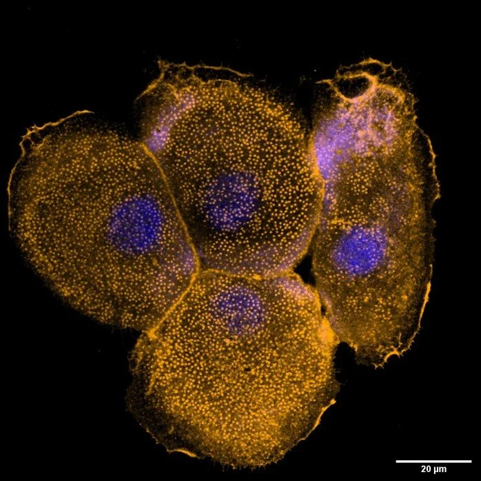
Human Keratinocyte on advanced 2D protein-patterned surface

Murine Embryonic Stem Cells (mESCs) micro topographies

(high-throughput imaging)