Morphology & Phenotyping
-
![]()
mESCs Phenotyping
-
![]()
Neural Stem Cell Phenotype
-
![]()
mESCs Fate Determination
-
![]()
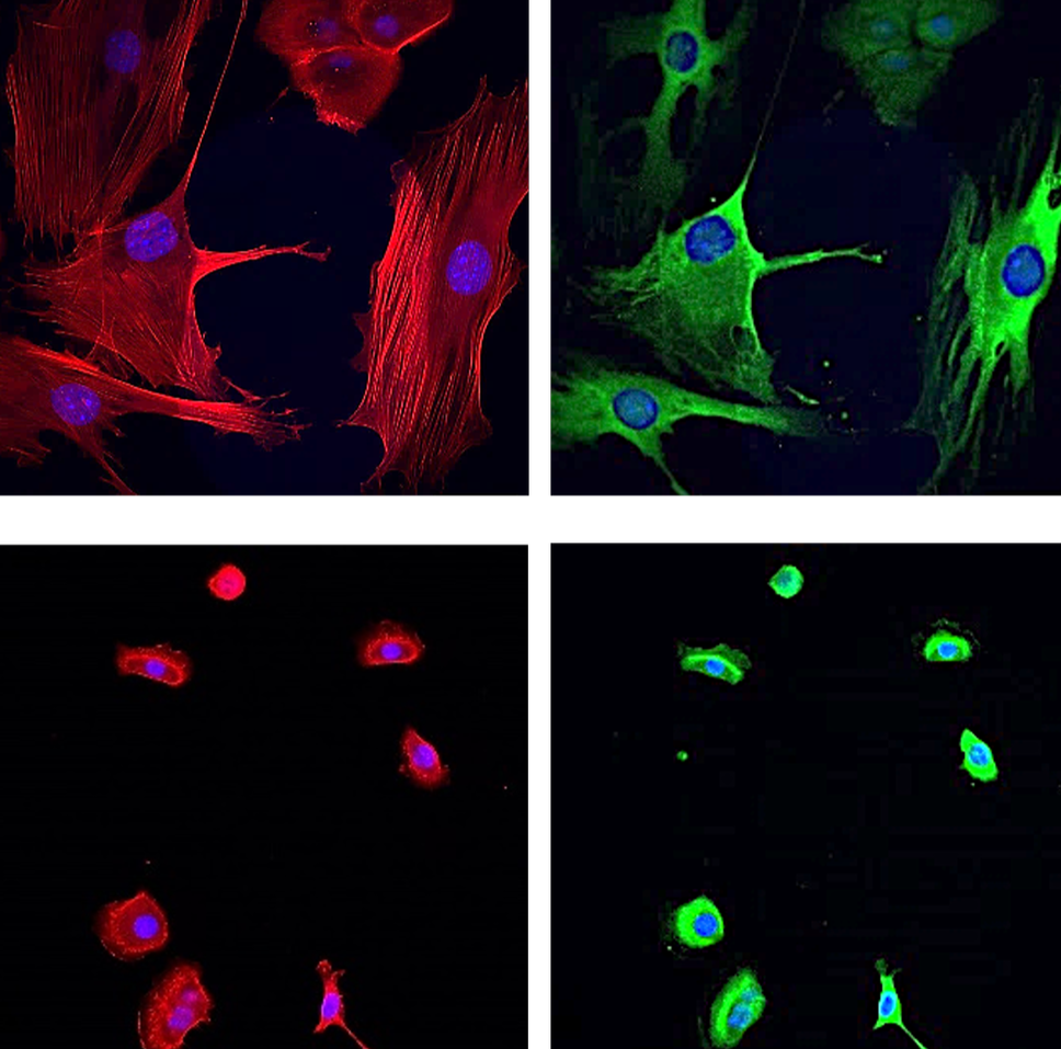
Epidermal Stem Cell Phenotype
Case Study 1: ECM-Driven Stemness and Morphological Phenotyping in mESCs
Overview
Assessment of how extracellular matrix (ECM) composition and media supplementation (LIF) influence stemness and colony morphology in murine embryonic stem cells (mESCs). Fluorescence imaging was used to evaluate OCT4 expression and structural organization across multiple ECM conditions.
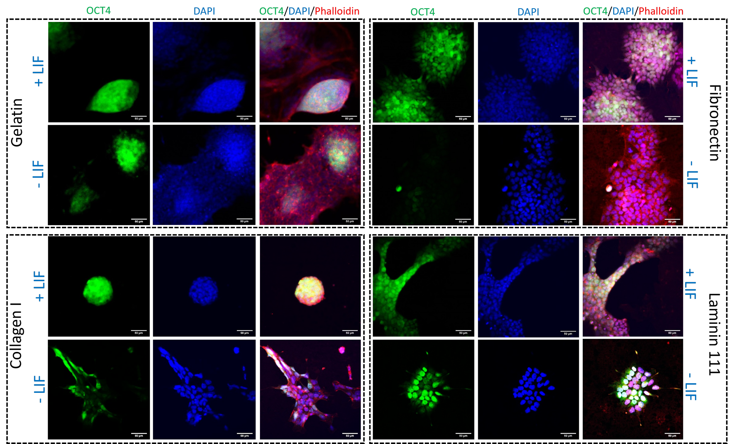
Fluorescence imaging of murine embryonic stem cells expressing an OCT4-GFP reporter cultured on various ECM substrates (gelatin, fibronectin, collagen I, and laminin-111) under +LIF and −LIF conditions. Nuclei are stained with DAPI (blue) and the actin cytoskeleton with phalloidin (red). Distinct ECM- and media-dependent changes in stemness and colony morphology are evident after 5 days of culture.

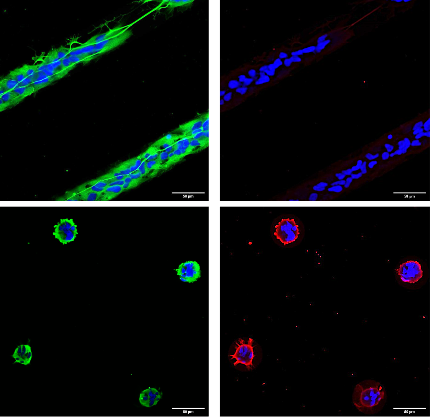
Case Study 2: Spatial Control of Neural Stem Cell Phenotype on ECM Micropatterns
Overview
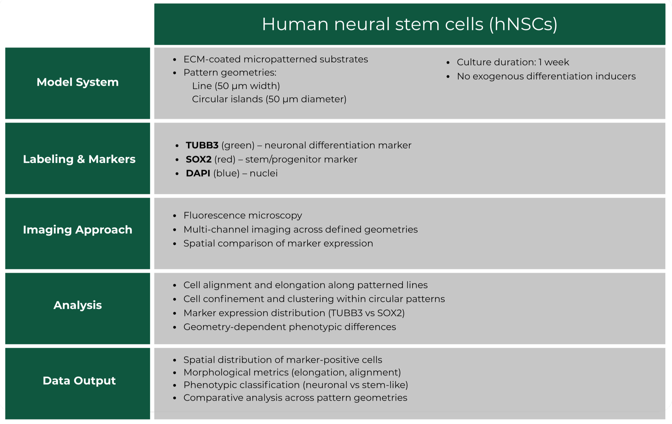
Investigation of how ECM micropattern geometry influences the morphology and phenotypic state of human neural stem cells (hNSCs). Fluorescence imaging was used to assess lineage-associated markers and the spatial organization of cells cultured on defined protein patterns in the absence of external differentiation cues.
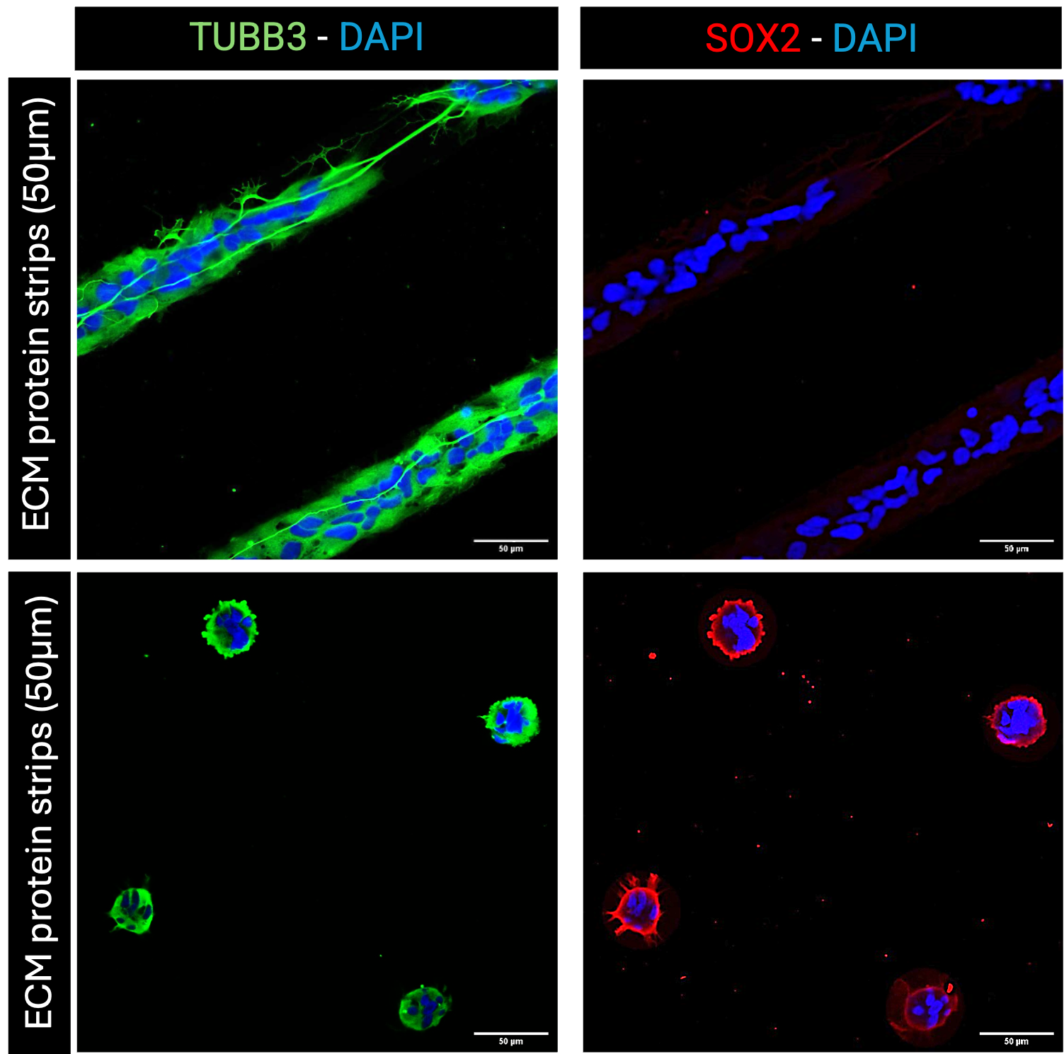
Fluorescence imaging of human neural stem cells cultured on ECM micropatterned substrates (50 μm line and circular geometries) for 1 week in the absence of differentiation inducers. Neuronal marker TUBB3 (green), stem cell marker SOX2 (red), and nuclei (DAPI, blue) reveal geometry-dependent differences in cell organization and phenotype. Line patterns promote aligned, elongated morphologies with increased neuronal features, while circular patterns maintain confined, stem-like cell states.
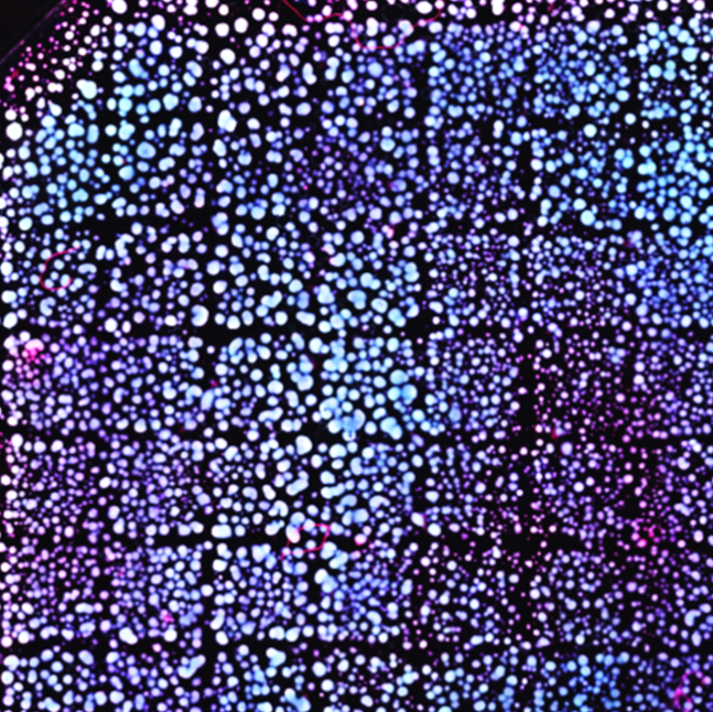
Case Study 3: Microenvironment-driven Fate Determination of mESCs on ECM-coated Submicron Topographies
Overview
Investigation of how extracellular matrix (ECM) composition and submicron-scale surface topographies regulate fate determination and spatial organization of murine embryonic stem cells (mESCs). High-content fluorescence imaging was used to assess stemness and morphological responses across combinatorial microenvironmental conditions.
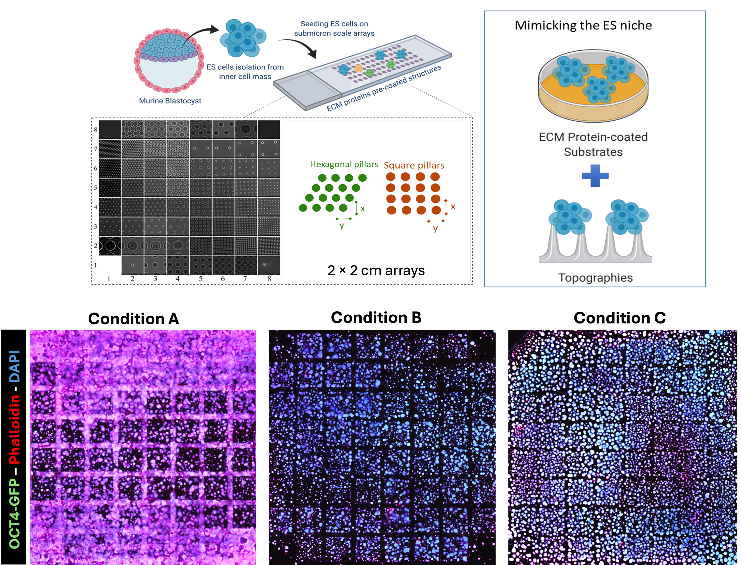
High-throughput fluorescence imaging of murine embryonic stem cells cultured on 2 × 2 cm submicron-patterned arrays comprising 64 distinct structures, pre-coated with different ECM ligand conditions (Condition A, Condition B, and controls), at day 5 post-seeding. OCT4 expression is visualized via a GFP reporter (green), with nuclei labeled by DAPI (blue) and F-actin by phalloidin (red). Multiparametric analysis reveals condition-dependent differences in cell distribution, morphology, and stemness across the patterned array.

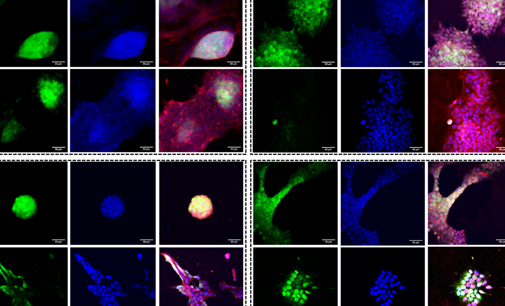
Case Study 4: Microenvironment and Calcium-dependent Regulation of Human Epidermal Stem Cell Phenotype
Overview
Investigation of how extracellular calcium levels and substrate architecture regulate the morphology and phenotypic state of human epidermal interfollicular stem cells (IFSCs). Fluorescence imaging was used to assess cytoskeletal organization and marker distribution across homogeneous and nanopatterned ECM environments.
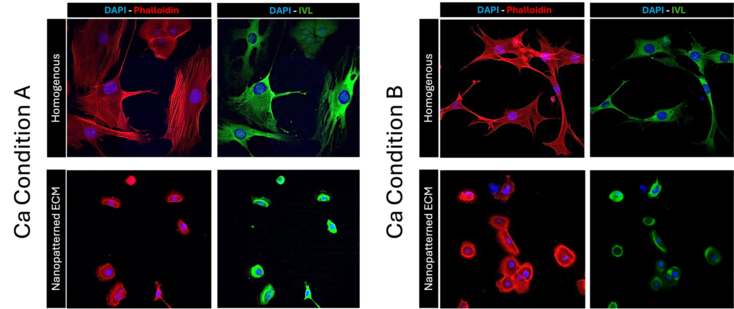
Fluorescence imaging of human epidermal interfollicular stem cells cultured on homogeneous and nanopatterned ECM substrates under defined calcium conditions (Condition A and Condition B). Cells are labeled for nuclei (DAPI, blue), actin cytoskeleton (phalloidin, red), and differentiation marker involucrin (IVL, green). Distinct differences in cell morphology, cytoskeletal organization, and phenotypic state are observed across substrate and calcium conditions.