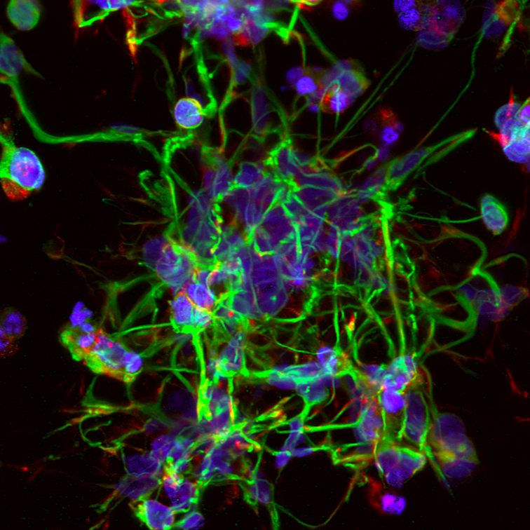
Human neural progenitor cells (NPCs)
Sample type
Human neural progenitor cells (NPCs) cultured in a 3D protein-engineered hydrogel microenvironment designed to mimic key biochemical and biomechanical features of brain extracellular matrix.
Cell source
Human iPSC-derived neural progenitor cells (human-relevant model).Culture format
Encapsulated NPCs in 3D hydrogelsStaining / markers (ICC/IF)
🟢 βIII-Tubulin (TUBB3): neuronal differentiation and neurite extension
🔴 F-actin (phalloidin): cytoskeletal organization and cell morphology
🔵 DAPI: nuclear identification
Microscopy & imaging
High-resolution confocal microscopy, visualization of cellular architecture within a 3D volume.
This image shows human neural progenitor cells cultured in a 3D, protein-engineered microenvironment, where immunofluorescence reveals neuronal differentiation, neurite outgrowth, and network formation at single-cell resolution.
By preserving spatial organization and protein localization, IF uncovers structural and signaling information that bulk and non-spatial assays cannot detect, enabling biologically meaningful insight in physiologically relevant models.
Read more
Related publication:
Viscoelastic N-cadherin-like interactions maintain neural progenitor cell stemness within 3D matrices, M. S. Huang, B. L. LeSavage, S. Ghorbani, et al. Nature Communications, (2025)