
Fluorescence imaging for seeing how biology responds.
CellCircuit helps discovery and translational teams convert fluorescence images into quantitative phenomic evidence, capturing morphology, marker localization, spatial organization, cell-state transitions, and microenvironment-dependent response across 2D, 3D, multiculture, patterned, and engineered cellular models.
These studies help teams understand not only whether cells respond, but how they respond, revealing phenotypic shifts, spatial patterns, and subcellular organization that viability-only, bulk, or endpoint assays can overlook.
Cellular phenomics across engineered model systems.
Representative fluorescence imaging studies showing how cell state, morphology, marker localization, cell–cell organization, cell–matrix interaction, and microenvironment-dependent phenotype can be measured across defined experimental conditions.
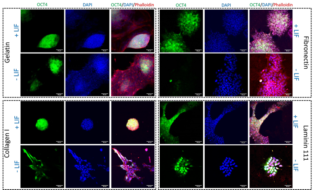
Fluorescence imaging of murine embryonic stem cells expressing OCT4-GFP on gelatin, fibronectin, collagen I, and laminin-111 under +LIF and −LIF conditions. DAPI labels nuclei and phalloidin labels F-actin, enabling comparison of pluripotency-associated signal, colony morphology, and cytoskeletal organization.
Stemness phenotyping across ECM-defined culture conditions
This study shows how extracellular matrix composition and media context can shift stemness-associated marker expression, colony structure, and cytoskeletal organization. For discovery teams, this type of assay helps evaluate whether culture conditions support the intended cell state before downstream screening or mechanistic studies.
Murine embryonic stem cells cultured on defined 2D ECM-coated substrates.
OCT4-GFP for pluripotency-associated state, DAPI for nuclei, and phalloidin for F-actin.
+LIF and −LIF media across gelatin, fibronectin, collagen I, and laminin-111.
Supports model-condition selection and early detection of phenotypic drift.
OCT4-GFP patterns across ECM and media conditions.
Compact, spread, or mixed colony phenotypes.
Actin architecture linked to colony structure and cell state.
Image-based comparison to guide model setup and assay conditions.
Related publication: Ghorbani S. et al., Biomaterials (2023).
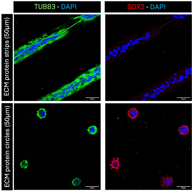
Fluorescence imaging of human neural stem/progenitor cells cultured on ECM micropatterned substrates with 50 μm line and circular geometries. TUBB3, SOX2, and DAPI reveal geometry-associated differences in morphology, alignment, confinement, and neural phenotype.
Geometry-guided phenotype control in neural stem/progenitor cells
This study demonstrates how defined spatial cues can influence neural stem/progenitor cell organization and lineage-associated marker patterns even without added differentiation factors. For CNS model development and phenotypic screening, this helps separate compound-driven effects from geometry- or culture-driven changes in cell state.
Human neural stem/progenitor cells cultured on ECM-coated line and circular micropatterns.
TUBB3 for neuronal-associated features, SOX2 for progenitor state, and DAPI for nuclei.
One-week culture on defined 50 μm geometries without exogenous differentiation inducers.
Guides assay geometry selection for controlled neural organization and phenotype.
Elongation, alignment, confinement, and clustering.
TUBB3 and SOX2 patterns across geometry conditions.
Marker-positive cells across line and circular ECM patterns.
Geometry selection for reproducible neural assay behavior.
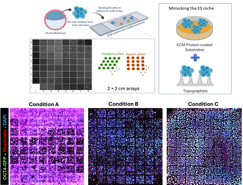
High-content fluorescence imaging of murine embryonic stem cells cultured on 2 × 2 cm submicron-patterned arrays with 64 structures, ECM ligand conditions, and controls. OCT4-GFP, DAPI, and phalloidin reveal differences in cell distribution, morphology, cytoskeletal organization, and stemness-associated signal across patterned environments.
Biomechanical cue-driven fate response on ECM-coated topographies
This study highlights how combinatorial microenvironmental conditions can be screened using fluorescence imaging and patterned arrays. For R&D teams, this approach helps identify substrate, ECM, and architecture-dependent phenotypes that influence assay reliability, cell fate, and interpretation of perturbation responses.
mESCs cultured on engineered submicron topographical arrays with multiple geometries.
OCT4-GFP for stemness-associated signal, DAPI for nuclei, and phalloidin for F-actin.
ECM-coated patterned substrates across ligand conditions and control environments.
Prioritizes microenvironmental conditions that produce desired or stable phenotypes.
Cell location and density across patterned array regions.
OCT4-GFP patterns across topography and ligand combinations.
Cell attachment and occupancy across engineered structures.
Structured morphology and marker profiles across many conditions.
Related publication: Ghorbani S. et al., Biomaterials (2023).
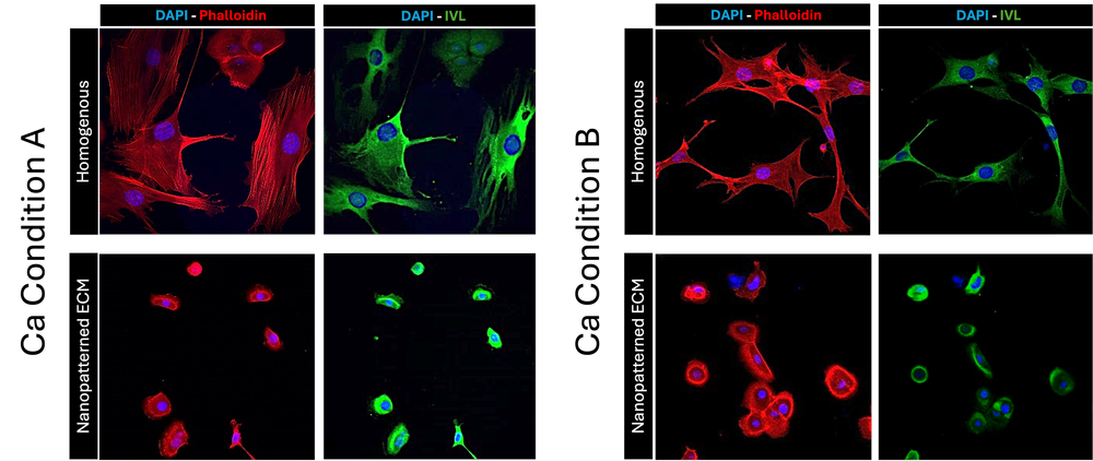
Fluorescence imaging of human epidermal interfollicular stem cells cultured on homogeneous and nanopatterned ECM substrates under defined calcium conditions. DAPI, phalloidin, and IVL reveal substrate- and calcium-dependent differences in morphology, cytoskeletal organization, and differentiation-associated phenotype.
Biochemical cue-driven fate response in epithelial stem cells
This study shows how biochemical cues and nanoscale substrate organization can jointly regulate epithelial stem cell morphology and differentiation-associated marker expression. These assays are relevant for epithelial biology, skin models, toxicity studies, wound-healing research, regenerative medicine, and phenotype-sensitive assay development.
Human epidermal interfollicular stem cells on flat ECM-coated and nanopatterned substrates.
DAPI for nuclei, phalloidin for F-actin, and IVL for involucrin expression.
Defined calcium levels across homogeneous and nanopatterned ECM environments.
Connects substrate design and media conditions to epithelial phenotype quality.
Cell area, spreading, elongation, and shape descriptors.
Actin organization across calcium and substrate conditions.
IVL-positive cells and differentiation-associated patterns.
Comparative imaging across flat and nanopatterned ECM systems.
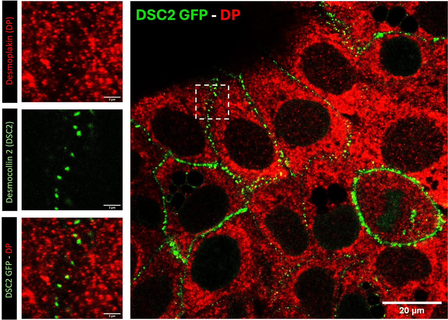
Desmoplakin, DP, red, and desmocollin-2, DSC2-GFP, green, localize at cell-cell junctions in HaCaT keratinocytes. Insets show protein clustering and co-localization at junctional regions.
Cell–cell interaction phenomics in epithelial monolayers
This study captures subcellular organization of desmosomal proteins at epithelial cell-cell junctions. For teams studying epithelial integrity, barrier models, adhesion biology, or compound-induced junctional remodeling, this type of imaging provides spatial evidence that complements marker intensity or bulk expression readouts.
2D epithelial monolayer culture of immortalized human keratinocytes with defined cell-cell junction formation.
Desmoplakin marks desmosomal plaques; DSC2-GFP reports desmosomal adhesion and localization.
High-resolution multi-channel fluorescence microscopy for subcellular junctional organization.
Supports evaluation of epithelial organization, junction integrity, and marker localization quality.
Localization of desmosomal proteins at cell-cell interfaces.
Overlap and distribution of DSC2 and desmoplakin signals.
Subcellular organization along epithelial cell boundaries.
Spatial evidence for junction formation and remodeling.
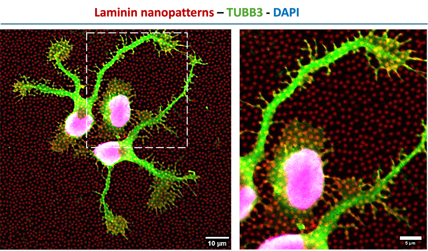
Human neural stem/progenitor cells on laminin-based ECM nanopatterns. TUBB3, green, marks neuronal-associated phenotype; ECM nanopatterns, red, define the engineered microenvironment; DAPI, magenta, labels nuclei.
Cell–ECM interaction phenomics in neural stem cells
This study shows how nanoscale ECM presentation can influence early neural morphology, marker distribution, and cell–matrix interaction. For teams developing neural models, this helps assess whether engineered substrates are guiding reproducible cellular organization and biologically relevant phenotypic behavior.
Human neural stem/progenitor cells cultured on laminin-based ECM nanopatterned substrates.
TUBB3 for neuronal-associated features, ECM pattern signal for substrate features, and DAPI for nuclei.
High-resolution multi-channel fluorescence imaging of cells, ECM patterns, and cell–matrix interactions.
Evaluates whether engineered ECM presentation supports desired neural organization and morphology.
Neurite extension, branching, and early neuronal-associated signatures.
Localized alignment and interaction with nanoscale pattern features.
Cell–ECM interaction maps and marker distribution.
Evidence to guide engineered microenvironment design.
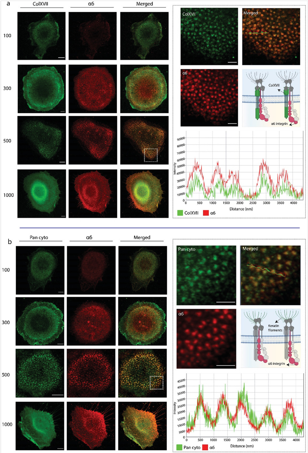
Confocal fluorescence imaging of HaCaT cells cultured on nanopatterned substrates with 100–1000 nm feature sizes. Collagen XVII, pan-cytokeratin, and α6 integrin reveal nanopattern-dependent adhesion and cytoskeletal organization.
Multi-biomarker co-localization on nanopatterned substrates
This study demonstrates how engineered nanoscale substrate features can affect adhesion-related marker localization and cytoskeletal organization. For epithelial model development, biomaterial screening, or phenotype-sensitive assay design, this type of analysis helps connect surface architecture to measurable cellular organization.
HaCaT keratinocytes cultured on nanopatterned substrates with defined 100–1000 nm feature sizes.
Collagen XVII and pan-cytokeratin paired with α6 integrin to assess adhesion and cytoskeletal organization.
Confocal laser scanning microscopy with multi-channel fluorescence imaging across nanopattern conditions.
Supports biomaterial and surface-feature selection for controlled epithelial phenotype.
Spatial correlation between adhesion and cytoskeletal markers.
Signal distribution across regions and nanopattern geometries.
Protein arrangement across nanoscale feature sizes.
Phenomic evidence for selecting engineered substrate conditions.
Related publication: Ghorbani S. et al., Small Methods (2022).